Several New Additions to the Scientific Database
Dec 1, 2016
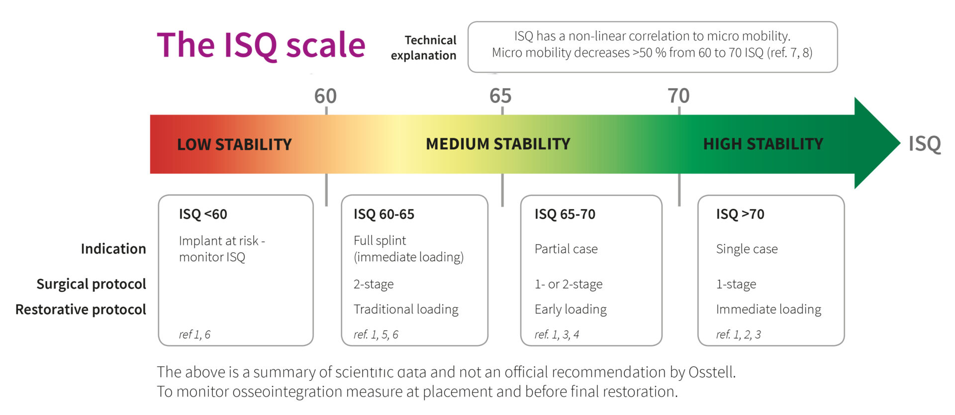
Our regularly updated database now includes more than 800 articles relating to Osstell and the ISQ scale. Below are a few of the studies that were recently published.
J Oral Maxillofac Surg. 2016 Jun; 74
The Predictive Value of Resonance Frequency Analysis Measurements in the Surgical Placement and Loading of Endosseous Implants
Baltayan S(1), Pi-Anfruns J(2), Aghaloo T(3), Moy PK(4)
PURPOSE: The purpose of this study was to investigate the predictive value of resonance frequency analysis in assessing implant survival. This was accomplished by determining the correlation between implant stability quotients (ISQs) and implant survival following different placement staging (1-stage vs 2-stage) and loading (early vs traditional) protocols.
MATERIALS AND METHODS: A retrospective study was performed on implant patient data collected over a 5-year period. Patients ranged in age from 16 to 91 years. We analyzed 703 implants during placement and 1,254 implants before loading. All implants were placed with respective ISQs recorded by 1 oral and maxillofacial surgeon. Receiver operating characteristic (ROC) statistical analysis was used to calculate sensitivity and specificity values corresponding to various ISQ cutoff points for different placement staging and loading protocols; χ(2) tests were used to identify significant differences.
RESULTS: In predicting implant failure, sensitivity progressively increased and specificity decreased as ISQ cutoff values increased. All failures occurred at an ISQ less than 66 for the placement staging protocol and an ISQ less than 67 for the loading protocol. When ISQ values were below 60, higher survival rates were observed when implants were placed using a 2-stage rather than a 1-stage placement staging protocol (P < .05). The area under the ROC curve for placement staging was 0.80, and the area under the ROC curve for loading was 0.89. An implant survival rate of over 98% was achieved.
CONCLUSIONS: Resonance frequency analysis is a noninvasive technique used to measure the stability of implants and to help guide placement staging and loading protocols. This study showed that increasing ISQ values correlated with increased sensitivity in detecting implant failure. Given the high survival rates of dental implants, additional studies can further elucidate the relationship between ISQ values and survival rates.
J Oral Implantol. 2016 Aug 19
Effect of heavy smoking on dental implants placed in male patients posterior mandibles: a prospective clinical study.
Sun C(1), Zhao J(2), Chen J(3), Tao H(4).
The objective of this study was to evaluate the bone healing and peri-implant tissue response in heavy smokers receiving dental implants due to partially edentulous posterior mandibles. Forty-five ITI Straumann dental implants were placed into the partially edentulous posterior mandibles of 16 heavy smokers and 16 non-smokers. One implant in each patient was evaluated for implant stability after surgery and before loading, and for the modified plaque index (mPLI), modified sulcus bleeding index (mSBI), probing depth (PD), and marginal bone loss (MBL) after loading. Meanwhile, the osteogenic capability of jaw marrow samples collected from patients was evaluated via an in vitro mineralization test.
For both groups, the implant stability quotient (ISQ) initially decreased from the initial ISQ achieved immediately after surgery and then increased starting from 2 weeks post-surgery. However, at 3, 4, 6, and 8 weeks post-surgery, the ISQ differed significantly between non-smokers and heavy smokers. All implants achieved osseointegration without complications at least by the end of 12th week post-surgery. At 6 or 12 months post-loading, the MBL and PD were significantly higher in heavy smokers than in non-smokers, whereas the mSBI and mPLI did not differ significantly between the two groups. The 1-year cumulative success rate of implants was 100% for both groups. Within the limitations of present clinical study (such as small sample size and short study duration), which applied the loading at 3 months post-operation, heavy smoking did not affect the cumulative survival rate of dental implants placed at the posterior mandible in male patients, but heavy smoking did negatively affect bone healing around dental implants by decreasing the healing speed.
These results implied that it might be of importance to select the right time point to apply the implant loading for heavy smokers. In addition, heavy smoking promoted the loss of marginal bone and the further development of dental pockets. Further clinical studies with larger patient populations are warranted to confirm our findings over a longer study duration.
J Craniofac Surg. 2016 Jul; 27
Immediate Loading of Tapered Implants Placed in Postextraction Sockets and Healed Sites
Han CH(1), Mangano F, Mortellaro C, Park KB.
OBJECTIVE: The aim of the present study was to compare the survival, stability, and complications of immediately loaded implants placed in post-extraction sockets and healed sites.
METHODS: Over a 2-year period, all patients presenting with partial or complete edentulism of the maxilla and/or mandible (healed site group, at least 4 months of healing after tooth extraction) or in need of replacement of nonrecoverable failing teeth (postextraction group) were considered for inclusion in this study. Tapered implants featuring a nanostructured calcium-incorporated surface were placed and loaded immediately. The prosthetic restorations comprised single crowns, fixed partial dentures, and fixed full arches. Primary outcomes were implant survival, stability, and complications. Implant stability was assessed at placement and at each follow-up evaluation (1 week, 3 months, and 1 year after placement): implants with an insertion torque (IT) <45 N·cm and/or with an implant stability quotient (ISQ) <70 were considered failed for immediate loading. A statistical analysis was performed.
RESULTS: Thirty implants were placed in postextraction sockets of 17 patients, and 32 implants were placed in healed sites of 22 patients. There were no statistically significant differences in ISQ values between the 2 groups, at each assessment. In total, 60 implants (96.8%) had an IT ≥45 and an ISQ ≥70 at placement and at each follow-up control: all these implants were successfully loaded. Only 2 implants (1 in a postextraction socket and 1 in a healed site, 3.2%) could not achieve an IT ≥45 N·cm and/or an ISQ ≥70 at placement or over time: accordingly, these were considered failed for stability, as they could not be subjected to immediate loading. One of these 2 implants, in a healed site of a posterior maxilla, had to be removed, yielding an overall 1-year implant survival rate of 98.4%. No complications were reported. No significant differences were reported between the 2 groups with respect to implant failures and complications.
CONCLUSION: Immediately loaded implants placed in post-extraction sockets and healed sites had similar high survival and stability, with no reported complications. Further long-term studies on larger samples of patients are needed to confirm these results.
Int J Oral Maxillofac Implants. 2016 May-Jun; 31
Lateral Alveolar Ridge Expansion in the Anterior Maxilla Using Piezoelectric Surgery for Immediate Implant Placement.
Nguyen VG, von Krockow N, Weigl P, Depprich R
PURPOSE: The purpose of this study was to evaluate the efficacy of the ridge-splitting technique in the anterior maxilla, using piezoelectric surgery for immediate implant placement. Study outcomes were compared with those of implant placement in the same patients using the conventional drilling technique.
MATERIALS AND METHODS: Ten patients received a total of 22 implants in the anterior maxilla, 11 of which were placed using a ridge-splitting procedure (test group) and the other 11 using the conventional drilling procedure (control group). Ridge width (RW), crestal bone level (BL), and implant stability quotient (ISQ) were measured at different points in time. Data were analyzed and compared between the groups using analysis of variance (ANOVA) and paired-sample tests at a significance level of 5%.
RESULTS: For the test group, the gain in RW was not stable in time because at 6 months postoperatively, the RW lost some of the initial gain; however, the net gain was still significant. At 6 months postoperatively, BL was similar for both groups. The net bone loss on the mesial aspect and the average of the mesial and the distal measures did not differ significantly between both groups. ISQ values sharply increased at 3 months postoperatively in the test group. All implants met the modified Albrektsson criteria (1989) for success.
CONCLUSION: The results from this study support the efficacy and safety of ridge expansion using piezoelectric surgery for implant insertion in the anterior maxilla. The modest net gain in bone width suggests that additional hard and soft tissue augmentation may be necessary, especially in the esthetic zone. ISQ values suggest a minimum healing time of 3 months before loading the implants that have been inserted using this ridge-splitting protocol.
J Orofac Orthop. 2016 Jul; 77
Orthodontic mini-implant stability at different insertion depths : Sensitivity of three stability measurement methods.
Nienkemper M(1), Santel N(2), Hönscheid R(2), Drescher D(2).
OBJECTIVES: The purpose of this work was to evaluate the influence of insertion depth on the stability of orthodontic mini-implants. Sensitivity of three different methods to measure implant stability based on differences in insertion depth were determined.
METHODS: A total of 82 mini-implants (2 × 9 mm) were inserted into pelvic bone of Swabian Hall pigs. Each implant was inserted stepwise to depths of 4, 5, 6, 7, and 8 mm. At each of these depths, three different methods were used to measure implant stability, including maximum insertion torque (MIT), resonance frequency analysis (RFA), and Periotest(®). Differences between the recorded values were statistically analyzed and the methods tested for correlations.
RESULTS: Almost linear changes from each insertion depth were measured with the values of RFA [implant stability quotient (ISQ) values range from 1-100], which increased from 6.95 ± 2.85 ISQ at 4 mm to 34.63 ± 5.51 ISQ at 8 mm, and with those of Periotest(®) [periotest values (PTV) range from -8 to 50], which decreased from 13.24 ± 4.03 PTV to -2.89 ± 1.87 PTV. Both methods were found to record highly significant (p < 0.0001) changes for each additional millimeter of insertion depth. The MIT increased significantly (p < 0.0001) from 153.67 ± 69.32 Nmm to 261 ± 103.73 Nmm between 4 and 5 mm of insertion depth but no further significant changes were observed as the implants were driven deeper. The RFA and Periotest(®) values were highly correlated (r = -0.907).
CONCLUSIONS: Mini-implant stability varies significantly with insertion depth. The RFA and the Periotest(®) yielded a linear relationship between stability and insertion depth. MIT does not appear to be an adequate method to determine implant stability based on insertion depth.
Clin Implant Dent Relat Res. 2016 Apr; 18
Influence of Skeletal and Local Bone Density on Dental Implant Stability in Patients with Osteoporosis.
Merheb J(1), Temmerman A(1), Rasmusson L(2), Kübler A(3), Thor A(4), Quirynen M(1,)(5).
BACKGROUND AND PURPOSE: Osteoporosis is a major skeletal disease affecting millions of people worldwide. Recent studies claim that patients with osteoporosis do not have a higher risk of early implant failure compared to non-osteoporotic patients. The aim of this study was to assess the effect of skeletal osteoporosis and local bone density on initial dental implant stability.
MATERIALS AND METHODS: Seventy-three patients were recruited and were assigned (based on a Dual-energy X-ray Absorptiometry scan) to either the osteoporosis (Opr), osteopenia (Opn), or control (C) group. Forty nine of the 73 patients received dental implants and had implant stability measured by means of resonance frequency analysis (RFA) at implant placement and at prosthetic abutment placement. On the computerized tomography scans, the cortical thickness and the bone density (Hounsfield Units) at the sites of implant placement were measured.
RESULTS: At implant placement, primary stability was on average lower in group Opr (63.3 ± 10.3 ISQ) than in group Opn (65.3 ± 7.5 implant stability quotient (ISQ), and group C (66.7 ± 8.7 ISQ). At abutment placement, a similar trend was observed: group Opr (66.4 ± 9.5 ISQ) scored lower than group Opn (70.7 ± 7.8 ISQ), while the highest average was for group C (72.2 ± 7.2 ISQ). The difference between groups Opr and C was significant. Implant length and diameter did not have a significant effect on implant stability as measured with RFA. A significant correlation was found between local bone density and implant stability for all regions of interest.
CONCLUSIONS: Implant stability seems to be influenced by both local and skeletal bone densities. The lower stability scores in patient with skeletal osteoporosis reinforce the recommendations that safe protocols and longer healing times could be recommended when treating those patients with dental implants.


